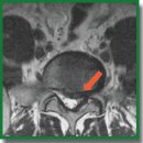
Diffusion-Weighted Magnetic Resonance Imaging in Diagnostics of Spinal Nerve Root Compression in Patients with Lumbar Intervertebral Disc Herniation
The aim of the study was to evaluate possibilities of using diffusion-weighted MRI (DW MRI) in diagnostics of compression of dorsal spinal ganglia and spinal nerve roots in patients with lumbar intervertebral disc herniation (IVD).
Materials and Methods. The study involved 37 patients (19 males, 18 females, average age — 42.6±12.7) with radicular syndrome caused by an IVD hernia of the lumbosacral spine. In all the cases, the diagnosis was confirmed by a clinical-neurological examination of a patient, radiography of the lumbar spine, multi-layer spiral CT and MRI investigations (including DW MRI). The control group included 29 volunteers (16 males, 13 females, average age — 37.4±9.6), who do not have clinical neurological and neuroimaging signs of a degenerative disease of lumbar IVD.
Results. Average value of the measured diffusion coefficient (MDC) compressed by the hernia of L5 IVD root amounted to 1634.7 mm2/s, and the value of the intact — to 1109.2 mm2/s. MDC for the right-sided S1 root without compression signs was 1392.5 mm2/s, while for the left-sided one it was 1375.7 mm2/s. The average MDC value for proximal regions of the spinal roots on the hernia side was 1441.2±13.7 mm2/s, and for the roots on the intact side it was 1243.9±17.6 mm2/s (р<0.001). Average MDC values for the distal regions of the roots on the degenerated IVD side were 1446.8±173.4 and 1107.5±76.1 mm2/s, respectively (р<0.001). There is an evident direct correlation between MDC of the dorsal spinal ganglion and pain intensity according to VAS (rs=0.089; p=0.012).
Conclusion. A specificity of compressed dorsal spinal ganglia and spinal nerve roots is their high MDC values which show microstructural changes manifested by edema, demyelination, and axonal injury. DW MRI, which enables the MDC to be calculated, is a perspective method of non-invasive instrumental diagnostics of compression nerve root syndromes in patients with degenerative IVD diseases.
- Mixter W.J., Barr J.S. Rupture of the intervertebral disc with involvement of the spinal canal. N Engl J Med 1934; 211: 210–215, https://doi.org/10.1056/nejm193408022110506.
- Krames E.S. The dorsal root ganglion in chronic pain and as a target for neuromodulation: a review. Neuromodulation 2015; 18(1): 24–32, https://doi.org/10.1111/ner.12247.
- Forget P., Boyer T., Steyaert A., Masquelier E., Deumens R., Le Polain de Waroux B. Clinical evidence for dorsal root ganglion stimulation in the treatment of chronic neuropathic pain. A review. Acta Anaesthesiol Belg 2015; 66(2): 37–41.
- Byvaltsev V.A., Stepanov I.A., Kalinin A.A., Shashkov K.V. Diffusion-weighted magnetic resonance tomography in the diagnosis of intervertebral disk degeneration. Biomedical Engineering 2016; 50(4): 253–256, https://doi.org/10.1007/s10527-016-9632-0.
- Byval’tsev V.A., Stepanov I.A., Kalinin A.A., Belykh E.G. Diffusion-weighted magnetic resonance imaging in the diagnosis of intervertebral disc degeneration in the lumbosacral spine. Vestnik rentgenologii i radiologii 2016; 97(6): 357–364.
- Byvaltsev V.A., Stepanov I.A., Kalinin A.A., Belykh E.G. Quantitative assessment of the degree of degenerative change in intervertebral disks using diffusion-weighted images. Biomedical Engineering 2017; 51(4): 275–279, https://doi.org/10.1007/s10527-017-9730-7.
- Byvaltsev V.A., Kolesnikov S.I., Belykh E.G., Stepanov I.A., Kalinin A.A., Bardonova L.A., Sudakov N.P., Klimenkov I.V., Nikiforov S.B., Semenov A.V., Perfil’ev D.V., Bespyatykh I.V., Antipina S.L., Giers M., Prul M. Complex analysis of diffusion transport and microstructure of an intervertebral disk. Bull Exp Biol Med 2017; 164(2): 223–228, https://doi.org/10.1007/s10517-017-3963-z.
- Byval’tsev V.A., Stepanov I.A., Semenov A.V., Perfil’ev D.V., Belykh E.G., Bardonova L.A., Nikiforov S.B., Sudakov N.P., Bespyatykh I.V., Antipina S.L. The possibilities for diagnostics of prescription of death coming based on the changes in the lumbar intervertebral disks (the comparison of the morphological, immunohistochemical and topographical findings). Sudebno-meditsinskaya ekspertiza 2017; 60(4): 4–8.
- Belykh E., Kalinin A.A., Patel A.A., Miller E.J., Bohl M.A., Stepanov I.A., Bardonova L.A., Kerimbaev T., Asantsev A.O., Giers M.B., Preul M.C., Byvaltsev V.A. Apparent diffusion coefficient maps in the assessment of surgical patients with lumbar spine degeneration. PLoS One 2017; 12(8): e0183697, https://doi.org/10.1371/journal.pone.0183697.
- Byvaltsev V.A., Stepanov I.A., Kalinin A.A., Belykh E.G. The use of apparent diffusion coefficient in diagnosis of lumbar intervertebral disk degeneration in patients with middle and old age by diffusion-weighted MRI. Uspekhi gerontologii 2018; 31(1): 103–109.
- Ohgiya Y., Oka M., Hiwatashi A., Liu X., Kakimoto N., Westesson P.A., Sven E., Ekholm S. Diffusion tensor MR imaging of the cervical spinal cord in patients with multiple sclerosis. Eur Radiol 2007; 17(10): 2499–2504, https://doi.org/10.1007/s00330-007-0672-4.
- Hiltunen J., Suortti T., Arvela S., Seppa M., Joensuu R., Hari R. Diffusion tensor imaging and tractography of distal peripheral nerves at 3 T. Clin Neurophysiol 2005; 116: 2315–2323, https://doi.org/10.1016/j.clinph.2005.05.014.
- Martín Noguerol T., Barousse R., Socolovsky M., Luna A. Quantitative magnetic resonance (MR) neurography for evaluation of peripheral nerves and plexus injuries. Quant Imaging Med Surg 2017; 7(4): 398–421, https://doi.org/10.21037/qims.2017.08.01.
- Yamashita T., Kwee T.C., Takahara T. Whole-body magnetic resonance neurography. N Engl J Med 2009; 361(5): 538–539, https://doi.org/10.1056/nejmc0902318.
- Takahara T., Imai Y., Yamashita T., Yasuda S., Nasu S., Van Cauteren M. Diffusion weighted whole body imaging with background body signal suppression (DWIBS): technical improvement using free breathing, STIR and high resolution 3D display. Radiat Med 2004; 22(4): 275–282.
- Eguchi Y., Ohtori S., Yamashita M., Yamauchi K., Suzuki M., Orita S., Kamoda H., Arai G., Ishikawa T., Miyagi M., Ochiai N., Kishida S., Inoue G., Masuda Y., Ochi S., Kikawa T., Toyone T., Takaso M., Aoki Y., Takahashi K. Diffusion-weighted magnetic resonance imaging of symptomatic nerve root of patients with lumbar disk herniation. Neuroradiology 2011; 53(9): 633–641, https://doi.org/10.1007/s00234-010-0801-7.
- Williams J.R. The declaration of Helsinki and public health. Bull World Health Organ 2008; 86(8): 650–652, https://doi.org/10.2471/blt.08.050955.
- Blagodatskiy M.D., Meyerovich S.I. Diagnostika i lechenie diskogennogo poyasnichno-kresttsovogo radikulita [Diagnosis and treatment of discogenic lumbosacral radiculitis]. Irkutsk: Izd-vo Irkutskogo un-ta; 1987; 271 p.
- Olmarker K., Rydevik B., Holm S. Edema formation in spinal nerve roots induced by experimental, graded compression: an experimental study on the pig cauda equina with special reference to differences in effects between rapid and slow onset of compression. Spine 1989; 14(6): 569–573, https://doi.org/10.1097/00007632-198906000-00003.
- Xue F., Wei Y., Chen Y., Wang Y., Gao L. A rat model for chronic spinal nerve root compression. Eur Spine J 2014; 23(2): 435–446, https://doi.org/10.1007/s00586-013-2990-3.
- Takata K., Inoue S., Takahashi K., Ohtsuka Y. Swelling of the cauda equina in patients who have herniation of a lumbar disc. A possible pathogenesis of sciatica. J Bone Joint Surg Am 1988; 70(3): 361–368, https://doi.org/10.2106/00004623-198870030-00007.
- Toyone T., Takahashi K., Kitahara H., Yamagata M., Murakami M., Moriya H. Visualisation of symptomatic nerve roots. Prospective study of contrast-enhanced MRI in patients with lumbar disc herniation. J Bone Joint Surg Br 1993; 75(4): 529–533, https://doi.org/10.1302/0301-620x.75b4.8331104.
- Germon T., Singleton W., Hobart J. Is NICE guidance for identifying lumbar nerve root compression misguided? Eur Spine J 2014; 23(S1): 20–24, https://doi.org/10.1007/s00586-014-3233-y.
- Lane J.I., Koeller K.K., Atkinson J.L. Contrast-enhanced radicular veins on MR of the lumbar spine in an asymptomatic study group. AJNR Am J Neuroradiol 1995; 16(2): 269–273.
- Boden S.D., Davis D.O., Dina T.S., Parker C.P., O’Malley S., Sunner J.L., Wiesel S.W. Contrast-enhanced MR imaging performed after successful lumbar disk surgery: prospective study. Radiology 1992; 182(1): 59–64, https://doi.org/10.1148/radiology.182.1.1727310.
- Park C.-K., Lee H.-J., Ryu K.-S. Comparison of root images between post-myelographic computed tomography and magnetic resonance imaging in patients with lumbar radiculopathy. Journal of Korean Neurosurgical Society 2017; 60(5): 540–549, https://doi.org/10.3340/jkns.2016.0809.008.
- Al-Tameemi H.N., Al-Essawi S., Shukri M., Naji F.K. Using magnetic resonance myelography to improve interobserver agreement in the evaluation of lumbar spinal canal stenosis and root compression. Asian Spine J 2017; 11(2): 198–203, https://doi.org/10.4184/asj.2017.11.2.198.
- Baliyan V., Das C.J., Sharma R., Gupta A.K. Diffusion weighted imaging: technique and applications. World J Radiol 2016; 8(9): 785–798, https://doi.org/10.4329/wjr.v8.i9.785.
- Ding W.Q., Gu J.H., Yuan Y., Jin D.S. Stereoscopic display of the peripheral nerves at the elbow region based on MR diffusion tensor imaging with multiple post-processing methods. Iran J Radiol 2016; 13(1): e22144, https://doi.org/10.5812/iranjradiol.22144.


