Evaluation of the Accuracy of Standardized Uptake Values of 18F-Fluorodeoxyglucose in Lung Lesions Based on Phantom Studies
The aim of the study was to estimate the accuracy of standardized uptake values of 18F-fluorodeoxyglucose (18F-FDG) in lung lesions during positron emission tomography combined with computed tomography (PET/CT) imaging, based on phantom studies performed for different PET/CT scanners.
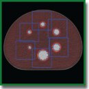
Materials and Methods. The analysis of the PET/CT with 18F-FDG data was performed for 86 patients newly diagnosed with the lung lesions: malignant tumors (n=37), benign tumors and inflammatory diseases (n=49). The criteria for inclusion in the study were developed considering the recommendations of the Fleischner Society (2017). The characteristics of the lesions on CT met the following requirements: a round shape or close to it; total size of 8 to 30 mm; solid or subsolid structure (with the exception of lesion with ground-glass opacity); a solid part size of ≥8 mm. All the patients had no signs of pleurisy, lymphadenopathy, or cancer history. PET/CT imaging with 18F-FDG was performed with three scanners: Discovery 690 (General Electric, USA), Biograph mCT 128 (Siemens, Germany), and Biograph mCT 40 (Siemens); the preparation of patients prior to the scan was standardized. To determine the reference accumulation of a radiopharmaceutical in the pathological lesion, four scans of a specialized NEMA IEC PET Body Phantom Set (USA) were performed for each scanner. For each unit, the recovery coefficients (RCs) of radioactivity, maximum and recovered (corrected) standardized uptake values (SUVs) were determined. Statistical relationship between the size of the lesion, SUVmax and SUVcorrect was evaluated. Data processing was performed using MedCalc v. 19.2.0 software.
Results. During the phantom study, the underestimation of the radioactivity was determined in the spheres with the diameters of 10 and 13 mm, overestimation was observed in the sphere with the diameter of 28 mm. Both underestimation and overestimation of radioactivity were determined for the spheres with a diameter of 17 and 22 mm.
SUVmax differed from the reference values for 85 patients (98.8%). The underestimation of these values was found for 63 patients (73.2%) due to the partial volume effect. The greatest underestimation was observed for the patients with 8 mm diameter lesions. Depending on the scanner, the underestimation of the SUVmax in these patients reached up to 54–73%. For 9 patients (25%) with malignant tumors of 9–12 mm, the utility of RC made it possible to avoid false negative results. For the lesions with a diameter of 30 mm, an overestimation of SUVmax up to 22% was determined due to the negative influence of the reconstruction algorithms.
Conclusion. The use of RC eliminates the influence of the partial volume effect and reconstruction methods on the accuracy of estimating the SUVmax in lung lesions, which ensures reproducibility, increase in the information content of the method, as well as the comparability of the results of PET/CT with 18F-FDG obtained on the different models of PET/CT units with different technological characteristics.
- Westerterp M., Pruim J., Oyen W., Hoekstra O., Paans A., Visser E., van Lanschot J., Sloof G., Boellaard R. Quantification of FDG PET studies using standardized uptake values in multi-center trials: effects of image reconstruction, resolution and ROI definition parameters. Eur J Nucl Med Mol 2007; 34(3): 392–404, https://doi.org/10.1007/s00259-006-0224-1.
- Lasnon C., Hicks R.J., Beauregard J.M., Milner A., Paciencia M., Guizard A.V., Bardet S., Gervais R., Lemoel G., Zalcman G., Aide N. Impact of point spread function reconstruction on thoracic lymph node staging with 18F-FDG PET/CT in non-small cell lung cancer. Clin Nucl Med 2012; 37: 971–976, https://doi.org/10.1097/rlu.0b013e318251e3d1.
- Armstrong I.S., Kelly M.D., Williams H.A., Matthews J.C. Impact of point spread function modelling and time of flight on FDG uptake measurements in lung lesions using alternative filtering strategies. EJNMMI Phys 2014; 1(1): 99, https://doi.org/10.1186/s40658-014-0099-3.
- Lindström E., Sundin A., Trampal C., Lindsjö L., Ilan E., Danfors T., Antoni G., Sörensen J., Lubberink M. Evaluation of penalized-likelihood estimation reconstruction on a digital time-of-flight PET/CT scanner for 18F-FDG whole-body examinations. J Nucl Med 2018; 59(7): 1152–1158, https://doi.org/10.2967/jnumed.117.200790.
- Bettinardi V., Castiglioni I., De Bernardi E., Gilardi M.C. PET quantification: strategies for partial volume correction. Clin Transl Imaging 2014; 2: 199–218, https://doi.org/10.1007/s40336-014-0066-y.
- Soret M., Bacharach S.L., Buvat I. Partial-volume effect in PET tumor imaging. J Nucl Med 2007; 8(6): 932–945, https://doi.org/10.2967/jnumed.106.035774.
- Hofeinz F., Langner J., Petr J., Beuthien-Baumann B., Oehme L., Steinbach J., Kotzerke J., van den Hoff J. A method for model-free partial volume correction in oncological PET. EJNMMI Res 2012; 2(1): 16, https://doi.org/10.1186/2191-219x-2-16.
- NEMA Standards Publication. NEMA NU2-2018. Performance measurement for Positron Emission Tomographs (PET). VA, USA: National Electrical Manufacturer Association; 2018.
- Pasawang P., Sontrapornpol T., Krisanachinda A. Experience on performance measurements of positron emission tomographs: NEMA NU2-2018. Med Phys Int 2019; 7(3): 305–313.
- Kaalep A., Sera T., Oyen W., Krause B.J., Chiti A., Liu Y., Boellaard R. EANM/EARL FDG-PET/CT accreditation — summary results from the first 200 accredited imaging systems. Eur J Nucl Med Mol Imaging 2018; 45(3): 412–422, https://doi.org/10.1007/s00259-017-3853-7.
- Rospotrebnadzor. Metodicheskie ukazaniya MUK 2.6.7.3651-20 “Metody kontrolya v PET-diagnostike dlya optimizacii radiacionnoj zashchity” [Methodical guidelines MUK 2.6.7.3651-20 “Methods of control in PET diagnostics to optimize radiation protection”]. Moscow; 2020; p. 34.
- MacMahon H., Naidich D., Goo J., Lee K.C., Leung A.N.C., Mayo J.R., Mehta A.C., Ohno Y., Powell C.A., Prokop M., Rubin G.D., Schaefer-Prokop C.M., Travis W.D., Van Schil P., Bankier A.A. Guidelines for management of incidental pulmonary nodules detected on CT images: from the Fleischner Society 2017. Radiology 2017; 84(1): 228–243, https://doi.org/10.1148/radiol.2017161659.
- Mukhortova O.V., Aslanidi I.P., Ashrafyan L.A., Shurupova I.V., Derevyanko E.P., Katunina T.A., Alimardonov D.B., Ulyanova A.V. 18F-fluorodeoxyglucose positron emission tomography in cancer patients: a whole-body examination procedure. Opuholi zenskoj reproduktivnoj sistemy 2009; 3–4: 70–77.
- Yablonskiy P.K., Tlostanova M.S., Avetisyan A.O. Efficacy of PET with 18F-FDG in differential diagnosis of lung cancer by calculation of standardized uptake value and tumor/lung criterion. Vestnik Sankt-Peterburgskogo universiteta. Seria 11. Medicina 2012; 1: 157–164.
- Boellaard R., Delgado-Bolton R., Oyen W.J.G., Giammarile F., Tatsch K., Eschner W., Verzijlbergen F.J., Barrington S.F., Pike L.C., Weber W.A., Stroobants S., Delbeke D., Donohoe K.J., Holbrook S., Graham M.M., Testanera G., Hoekstra O.S., Zijlstra J., Visser E., Hoekstra C.J., Pruim J., Willemsen A., Arends B., Kotzerke J., Bockisch A., Beyer T., Chiti A., Krause B.J; European Association of Nuclear Medicine (EANM). FDG PET/CT: EANM procedure guidelines for tumour imaging: version 2.0. Eur J Nucl Med Mol Imaging 2015; 42(2): 328–354, https://doi.org/10.1007/s00259-014-2961-x.
- Sher A., Lacoeuille F., Fosse P., Vervueren L., Cahouet-Vannier A., Dabli D., Bouchet F., Couturier O. For avid glucose tumors, the SUV peak is the most reliable parameter for [18F]FDGPET/CT quantification, regardless of acquisition time. EJNMMI Res 2016; 6(1): 21, https://doi.org/10.1186/s13550-016-0177-8.
- Zhang Q., Gao X., Wei G., Qiu C., Qu H., Zhou X. Prognostic value of MTV, SUVmax and the T/N ratio of PET/CT in patients with glioma: a systematic review and meta-analysis. J Cancer 2019; 10(7): 1707–1716, https://doi.org/10.7150/jca.28605.
- Li Q., Zhang J., Cheng W., Zhu C., Chen L., Xia F., Wang M., Yang F., Ma X. Prognostic value of maximum standard uptake value, metabolic tumor volume, and total lesion glycolysis of positron emission tomography/computed tomography in patients with nasopharyngeal carcinoma: a systematic review and meta-analysis. Medicine 2017; 96(37): e8084, https://doi.org/10.1097/md.0000000000008084.
- Keyes J.W. Jr. SUV: standard uptake or silly useless value? J Nucl Med 1995; 36(10): 1836–1839.
- Sugawara Y., Zasadny K.R., Neuhoff A.W., Wahl R.L. Reevaluation of the standardized uptake value for FDG: variations with body weight and methods for correction. Radiology 1999; 213(2): 521–525, https://doi.org/10.1148/radiology.213.2.r99nv37521.
- Aide N., Lasnon C., Veit-Haibach P., Sera T., Sattler B., Boellaard R. EANM/EARL harmonization strategies in PET quantification: from daily practice to multicentre oncological studies. Eur J Nucl Med Mol Imaging 2017; 44 (Suppl 1): 17–31, https://doi.org/10.1007/s00259-017-3740-2.
- Hoetjes N.J., van Velden F.H., Hoekstra O.S., Hoekstra C.J., Krak N.C., Lammertsma A.A., Boellaard R. Partial volume correction strategies for quantitative FDG PET in oncology. Eur J Nucl Med Mol Imaging 2010; 37: 1679–1687, https://doi.org/10.1007/s00259-010-1472-7.
- Sonni I., Baratto L., Park S., Hatami N., Srinivas S., Davidzon G., Gambhir S.S., Iagaru A. Initial experience with a SiPM-based PET/CT scanner: influence of acquisition time on image quality. EJNMMI Phys 2018; 5(1): 9, https://doi.org/10.1186/s40658-018-0207-x.
- Wagatsuma K., Miwa K., Sakata M., Oda K., Ono H., Kameyama M., Toyohara J., Ishii K. Comparison between new-generation SiPM-based and conventional PMT-based TOF-PET/CT. Phys Med 2017; 42: 203–210, https://doi.org/10.1016/j.ejmp.2017.09.124.
- Granov A.M., Tyutin L.А., Tlostanova М.S., Avetisyan А.О., Ryzhkova D.V. Optimization of quantitative processing data of positron emission tomography with 18F-FDG in patients with lung cancer. Sovremennye tehnologii v medicine 2012; 1: 44–48.
- Tlostanova M.S., Avetisyan A.O. The informativeness of positron emission tomography with [18F]-fluorodeoxyglucose in differential diagnosis of lung cancer. Vestnik Rossiyskogo gosudarstvennogo meditsinskogo universiteta 2012; 2: 41–44.
- Khalaf M., Abdel-Nabi H., Baker J., Shao Y., Lamonica D., Gona J. Relation between nodule size and 18F-FDG-PET SUV for malignant and benign pulmonary nodules. J Hematol Oncol 2008; 1–8: 13, https://doi.org/10.1186/1756-8722-1-13.
- Yilmaz F., Tastekin G. Sensitivity of 18F-FDG PET in evaluation of solitary pulmonary nodules. Int J Clin Exp Med 2015; 8(1): 45–51.
- Christian P.E. Use of a precision fillable clinical simulator phantom for PET/CT scanner validation in multi-center clinical trials: the SNM Clinical Trials Network (CTN) Program. J Nucl Med 2012; 53(Suppl 1): 437.
- Lasnon С., Quak E., Le Roux P.Y., Robin F., Hofman M.S., Bourhis D., Callahan J., Binns D.S., Desmonts C., Salaun P.Y., Hicks R.J., Aide N. EORTC PET response criteria are more influenced by reconstruction inconsistencies than PERCIST but both benefit from the EARL harmonization program. EJNMMI Physics 2017; 4(1): 17, https://doi.org/10.1186/s40658-017-0185-4.
- Sunderland J.J., Christian P.E. Quantitative PET/CT scanner performance characterization based upon the society of nuclear medicine and molecular imaging clinical trials network oncology clinical simulator phantom. J Nucl Med 2015; 56(1): 145–152, https://doi.org/10.2967/jnumed.114.148056.


