Experimental Study of the Effect of Corneal Bio-Coating Based on the Original Hydrogel Biopolymer Scaffold on the Anterior Segment Structures of the Eye
In recent years, there have been proposed various kinds of bioengineering constructions of scaffolds — biomaterial structures serving as substrates for restoration and regeneration of tissues. They possess marked biocompatibility, are capable of supporting high concentration of substances delivered to the injured tissues, and may be standardized in the process of fabrication.
The aim of the investigation is to study experimentally the effect of the bio-coating based on the original hydrogel biopolymer scaffold on the intact structures of the anterior segment of the eye and its adnexa and evaluate its safety.
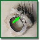
Materials and Methods. The experimental study was carried out on 6 rabbits (6 eyes). The original hydrogel biopolymer scaffold of 10.0 mm in diameter, 1.5 and 2.0 mm thick was used as a bio-coating for the cornea. On days 3, 7, and 9 from the beginning of the experiment, the results were histologically tested.
Results. On days 3 and 7 of the experiment, histological investigations did not find any structural changes of the anterior segment of the eye and differences between the scaffolds of various thicknesses. The stroma, Descemet’s membrane, endothelium, and general topography of the intercellular matrix were not altered. On day 9, structural changes of the anterior segment were not revealed either in the experiment with the 1.5 mm thick scaffold. Histological investigations of the specimen with the 2.0 mm scaffold showed alterations in the form of epithelium thickening, signs of pseudostratified basal layer, hyperplasia of the wing cell layer with the increased number of its layers, greater number of cellular elements in the anterior stroma layers. No structural changes of the Descemet’s membrane and corneal endothelium were noted.
Conclusion. The suggested hydrogel scaffold-based bio-coating is subject to self-biodegradation without any sequelae to the eye and its adnexa. Increased thickness of the bio-coating results in deceleration of its biodegradation and enhanced activity of proliferative processes in the epithelium and anterior stromal layers, which is indirect evidence of improved regenerative properties of these tissues.
- Burton M.J., Ramke J., Marques A.P., Bourne R.R.A., Congdon N., Jones I., Ah Tong B.A.M., Arunga S., Bachani D., Bascaran C., Bastawrous A., Blanchet K., Braithwaite T., Buchan J.C., Cairns J., Cama A., Chagunda M., Chuluunkhuu C., Cooper A., Crofts-Lawrence J., Dean W.H., Denniston A.K., Ehrlich J.R., Emerson P.M., Evans J.R., Frick K.D., Friedman D.S., Furtado J.M., Gichangi M.M., Gichuhi S., Gilbert S.S., Gurung R., Habtamu E., Holland P., Jonas J.B., Keane P.A., Keay L., Khanna R.C., Khaw P.T., Kuper H., Kyari F., Lansingh V.C., Mactaggart I., Mafwiri M.M., Mathenge W., McCormick I., Morjaria P., Mowatt L., Muirhead D., Murthy G.V.S., Mwangi N., Patel D.B., Peto T., Qureshi B.M., Salomão S.R., Sarah V., Shilio B.R., Solomon A.W., Swenor B.K., Taylor H.R., Wang N., Webson A., West S.K., Wong T.Y., Wormald R., Yasmin S., Yusufu M., Silva J.C., Resnikoff S., Ravilla T., Gilbert C.E., Foster A., Faal H.B. The Lancet Global Health Commission on Global Eye Health: vision beyond 2020. Lancet Glob Health 2021; 9(4): e489–e551, https://doi.org/10.1016/S2214-109X(20)30488-5.
- Neroev V.V. Rabota Rossiyskogo natsional’nogo komiteta po likvidatsii ustranimoy slepoty v ramkakh programmy VOZ “Zrenie 2020”. V kn.: Materialy VIII mezhregional’nogo simpoziuma “Profilaktika slepoty vsledstvie travm organa zreniya”. Initsiativa VOZ “Vseobshchiy dostup k zdorov’yu glaz: global’nyy plan deystviy na 2014–2019 gody”, 13 oktyabrya 2016 [The work of the Russian National Committee for the Elimination of Eradicable Blindness within the framework of the WHO Vision 2020 program. In: Proceedings of the VIII interregional symposium “Prevention of blindness due to eye injuries”. WHO initiative “Universal access to eye health: global action plan 2014–2019”, October 13, 2016]. Moscow; 2017; p. 11–34.
- Thia Z.Z., Ho Y.T., Shih K.C., Tong L. New developments in the management of persistent corneal epithelial defects. Surv Ophthalmol 2023; 68(6): 1093–1114, https://doi.org/10.1016/j.survophthal.2023.06.001.
- Trufanov S.V., Subbot A.M., Shakhbazyan N.P. Modern biotechnological treatment methods of persistent corneal epithelial defects. Vestn Oftalmol 2020; 136(5): 277–282, https://doi.org/10.17116/oftalma2020136052277.
- Bezushko A.V., Dubovikov A.S., Kulikov A.N., Churashov S.V., Chernysh V.F., Blinova M.I., Aleksandrova O.I., Khorolskaya Yu.I., Gavrilyu I.O., Karpovich V.V., Danilichev V.F. The use of collagen scaffold and amniotic membrane with laboratory-reared stem cells to manage limbal deficiency: experimental study. Tihookeanskij medicinskij zurnal 2019; 2: 54–57.
- Mazzocca A.D., McCarthy M.B., Chowaniec D.M., Dugdale E.M., Hansen D., Cote M.P., Bradley J.P., Romeo A.A., Arciero R.A., Beitzel K. The positive effects of different platelet-rich plasma methods on human muscle, bone, and tendon cells. Am J Sports Med 2012; 40(8): 1742–1749, https://doi.org/10.1177/0363546512452713.
- Fedoseeva E.V., Chentsova E.V., Borovkova N.V., Ponomarev I.N., Vlasova V.A., Pavlenko Yu.A. Experience of using a thrombofibrin clot of plateletrich plasma in ulcerative lesions of the cornea. Rossijskij oftalʹmologiceskij zurnal 2021; 14(4): 15–21, https://doi.org/10.21516/2072-0076-2021-14-4-supplement-15-21.
- You J., Hodge C., Hoque M., Petsoglou C., Sutton G. Human platelets and derived products in treating ocular surface diseases — a systematic review. Clin Ophthalmol 2020; 14: 3195–3210, https://doi.org/10.2147/OPTH.S265701.
- Natalia M.E.R., Susiyanti M. The efficacy of sutureless amnion membrane transplantation for corneal epithelialization in delayed corneal healing: a systematic review. Korean J Ophthalmol 2025; 39(3): 288–299, https://doi.org/10.3341/kjo.2025.0004.
- Novitsky I.Ya., Sarakhman M.N., Smal T.M. Transplantation of amniotic membrane with fixation in the corneal layers. Oftal’mokhirurgiya 2003; 3: 4–7.
- Furundaoturan O., Palamar M., Barut Selver Ö. Palliative efficacy of intrastromal amniotic membrane procedure in symptomatic bullous keratopathy patients. Turk J Ophthalmol 2022; 52(3): 162–167, https://doi.org/10.4274/tjo.galenos.2022.38839.
- Meller D., Pauklin M., Thomasen H., Westekemper H., Steuhl K.P. Amniotic membrane transplantation in the human eye. Dtsch Arztebl Int 2011; 108(14): 243–248, https://doi.org/10.3238/arztebl.2011.0243.
- Egorikhina M.N., Mukhina P.A., Bronnikova I.I. Scaffolds as drug and bioactive compound delivery systems. Kompleksnye problemy serdečno-sosudistyh zabolevanij 2020; 9(1): 92–102, https://doi.org/10.17802/2306-1278-2020-9-1-92-102.
- Egorikhina M.N., Levin G.Ya., Charykova I.N., Alejnik D.Ya., Sosnina L.N. Method for creating a bioresorbable cellular scaffold based on fibrin of blood plasma. Patent RU 2653434 C1.
- Semenycheva L.L., Astanina M.V., Kuznetsova J.L., Valetova N.B., Geras’kina E.V., Tarankova O.A. Method for production of acetic dispersion of high molecular fish collagen. Patent RU 2567171 C1.
- Egorikhina M.N., Aleynik D.Y., Rubtsova Y.P., Levin G.Y., Charykova I.N., Semenycheva L.L., Bugrova M.L., Zakharychev E.A. Hydrogel scaffolds based on blood plasma cryoprecipitate and collagen derived from various sources: structural, mechanical and biological characteristics. Bioact Mater 2019; 4: 334–345, https://doi.org/10.1016/j.bioactmat.2019.10.003.
- Matthews J.A., Wnek G.E., Simpson D.G., Bowlin G.L. Electrospinning of collagen nanofibers. Biomacromolecules 2002; 3(2): 232–238, https://doi.org/10.1021/bm015533u.
- Chen S., Mienaltowski M.J., Birk D.E. Regulation of corneal stroma extracellular matrix assembly. Exp Eye Res 2015; 133: 69–80, https://doi.org/10.1016/j.exer.2014.08.001.
- Vogel W.F. Collagen-receptor signaling in health and disease. Eur J Dermatol 2001; 11(6): 506–514.
- Baratta R.O., Del Buono B.J., Schlumpf E., Ceresa B.P., Calkins D.J. Collagen mimetic peptides promote corneal epithelial cell regeneration. Front Pharmacol 2021; 12: 705623, https://doi.org/10.3389/fphar.2021.705623.
- Chattopadhyay S., Raines R.T. Review collagen-based biomaterials for wound healing. Biopolymers 2014; 101(8): 821–833, https://doi.org/10.1002/bip.22486.
- Egorikhina M.N. The use of blood components in tissue engineering. Siberian Medical Review 2018; 3: 14–23, https://doi.org/10.20333/2500136-2018-3-14-23.
- Martino M.M., Briquez P.S., Ranga A., Lutolf M.P., Hubbell J.A. Heparin-binding domain of fibrin(ogen) binds growth factors and promotes tissue repair when incorporated within a synthetic matrix. Proc Natl Acad Sci U S A 2013; 110(12): 4563–4568, https://doi.org/10.1073/pnas.1221602110.
- Martino M.M., Hubbell J.A. The 12th–14th type III repeats of fibronectin function as a highly promiscuous growth factor-binding domain. FASEB J 2010; 24(12): 4711–4721, https://doi.org/10.1096/fj.09-151282.


