Laser Soldering with a Biosolder for Oral Mucosa Wound Closure in an Experiment
The aim of the study was to evaluate the efficiency of wound closure of soft tissues using a 970 nm diode laser and a biosolder based on bovine serum albumin and single-wall carbon nanotubes in experimentally modelled oral cavity defects.
Materials and Methods. The experimental study was carried out on 16 chinchilla rabbits. Linear defects of the oral mucosa 1 cm long were modeled. The animals were divided into 2 groups, 8 rabbits in each group. The wounds in the control group animals were sutured with a surgical suture using Prolene 5-0 thread; the experimental group animals were sutured using laser soldering and a biosolder based on bovine serum albumin, indocyanine green, single-wall carbon nanotubes, and type I collagen, followed by putting additional sutures using Prolene 5-0 thread. We used the proprietary laser device with a wavelength of 970 nm with adaptive thermal stabilization of the suture, which enabled to set the heating temperature of the biotissue in the laser suture area with an accuracy of ~1°C preventing thermal necrosis of tissues.
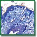
The biological tissues of 24 samples of the rabbit oral mucosa were fixed on days 1, 3, 5, and 10 and examined morphologically and morphometrically.
Results. Inflammatory changes were primarily associated with a response to the suture material; proliferative changes (neoangiogenesis and epithelial regeneration) were related to the proliferation activation of fibroblasts and epithelial cells due to the laser exposure. The use of a biosolder contributed to additional tissue adhesion, which further on accelerated the regeneration process and increased the neoangiogenesis rate and the vascular density per 1 mm2.
In the experimental group, the inflammatory reaction was completed by day 5, while in the control group, the residual inflammatory signs persisted in some samples up to day 10. On day 10, the proliferative phase began in the experimental group. An immunohistochemical analysis revealed a statistically significant increase in the number of blood vessels in the experimental group by 70.6% compared to the control (p=0.003).
Conclusion. The use of laser exposure combined with a biosolder promoted tissue adhesion improvement, shortened the inflammatory phase, and accelerated the regeneration providing minimal scarring. The data obtained emphasize the prospects of using the suggested technique for oral mucous wound closure in clinical practice for patients with various dental diseases.
- Faris A., Khalid L., Hashim M., Yaghi S., Magde T., Bouresly W., Hamdoon Z., Uthman A.T., Marei H., Al-Rawi N. Characteristics of suture materials used in oral surgery: systematic review. Int Dent J 2022; 72(3): 278–287, https://doi.org/10.1016/j.identj.2022.02.005.
- La Rosa G.R.M., Scapellato S., Cicciù M., Pedullà E. Antimicrobial activity of antibacterial sutures in oral surgery: a scoping review. Int Dent J 2024; 74(4): 688–695, https://doi.org/10.1016/j.identj.2024.01.029.
- Durnovo E.A., Tarakanova V.A. Opportunities of the wound process optimization using photodynamic therapy on the mucous membrane in the oral cavity. Dental Forum 2019; 4(75): 35–36.
- Tarasenko S.V., Blagushina N.A. Experimental histological evaluation of bioresorbable collagen membrane use in surgical oral mucosal defects. Vyatskiy meditsinskiy vestnik 2022; 1(73): 67–75.
- Sharipov I.A., Ditkovsky V.V., Khatomkin D.M., Komissarova N.V. Knots and sutures in surgery. Sinergiya nauk 2022; 71: 546–563.
- Larjava H., Wiebe C., Gallant-Behm C., Hart D.A., Heino J., Häkkinen L. Exploring scarless healing of oral soft tissues. J Can Dent Assoc 2011; 77: b18.
- Chandra G.B., VinayKumar M.B., Walavalkar N.N., Vandana K.L., Vardhan P.K. Evaluation of surgical scalpel versus semiconductor diode laser techniques in the management of gingival melanin hyperpigmentation: a split-mouth randomized clinical comparative study. J Indian Soc Periodontol 2020; 24(1): 47–53, https://doi.org/10.4103/jisp.jisp_186_19.
- Blashkova S.L., Krikun E.V., Mustafin I.G., Valeeva I.K., Blashkova J.V. Dynamics of clinical and immunological parameters in the integrated management of endoperiodontal lesions, including laser therapy. Kazanskiy meditsinskiy zhurnal 2021; 102(3): 322–328, https://doi.org/10.17816/KMJ2021-322.
- Gerasimenko A.Y., Morozova E.A., Ryabkin D.I., Fayzullin A., Tarasenko S.V., Molodykh V.V., Pyankov E.S., Savelyev M.S., Sorokina E.A., Rogalsky A.Y., Shekhter A., Telyshev D.V. Reconstruction of soft biological tissues using laser soldering technology with temperature control and biopolymer nanocomposites. Bioengineering (Basel) 2022; 9(6): 238, https://doi.org/10.3390/bioengineering9060238.
- Matteini P., Ratto F., Rossi F., de Angelis M., Cavigli L., Pini R. Hybrid nanocomposite films for laser-activated tissue bonding. J Biophotonics 2012; 5(11–12): 868–877, https://doi.org/10.1002/jbio.201200115.
- Ark M., Cosman P.H., Boughton P., Dunstan C.R. Photochemical tissue bonding (PTB) methods for sutureless tissue adhesion. Int J Adhes Adhes 2016; 71: 87–98, https://doi.org/10.1016/j.ijadhadh.2016.08.006.
- Judy M.M., Fuh L., Matthews J.L., Lewis D.E., Utecht R.E. Gel electrophoretic studies of photochemical cross-linking of type I collagen with brominated 1,8-naphthalimide dyes and visible light. Proc. SPIE 2128, Laser Surgery: Advanced Characterization, Therapeutics, and Systems IV 1994, https://doi.org/10.1117/12.184876.
- Judy M.M., Nosir H.R., Jackson R.W., Matthews J.L., Utecht R.E., Lewis D.E., Yuan D. Photochemical bonding of skin with 1,8-naphthalimide dyes. Proc. SPIE 3195, Laser-Tissue Interaction, Tissue Optics, and Laser Welding III 1998, https://doi.org/10.1117/12.297902.
- Merguerian P.A., Pugach J.L., Lilge L.D. Nonthermal ureteral tissue bonding: Nonthermal ureteral tissue bonding: comparison of photochemical collagen crosslinking with thermal laser bonding. Proc. SPIE 3590, Lasers in Surgery: Advanced Characterization, Therapeutics, and Systems IX 1999; https://doi.org/10.1117/12.350962.
- Mulroy L., Kim J., Wu I., Scharper P., Melki S.A., Azar D.T., Redmond R.W., Kochevar I.E. Photochemical keratodesmos for repair of lamellar corneal incisions. Invest Ophthalmol Vis Sci 2000; 41(11): 3335–3340.
- Bespalova N.A., Durnovo E.A., Galkina E.S., Tarakanova V.A., Runova N.V. The infrared thermometry: registration of healing process of the free gingival graft. Parodontologiya 2020; 25(2): 127–133, https://doi.org/10.33925/1683-3759-2020-25-2-127-133.
- Ashurko I.P., Krylova D.A., Belkin V.O., Yatsenko A.G., Tarasenko S.V. Results of using collagen matrix in soft tissue management in the area of dental implants in the anterior region of the upper jaw. Problemy stomatologii 2023; 19(4): 69–76, https://doi.org/10.18481/2077-7566-2023-19-4-69-76.
- Antoshin A., Gostev M., Khristidis Y., Giliazova A., Voloshin S., Blagushina N., Smirnova O., Diachkova E., Istranova E., Usanova A., Solodov N., Fayzullin A., Ivanova E., Sadchikova E., Vergara Bashkatova M.N., Drakina O., Tarasenko S., Timashev P. Electrophoretically co-deposited collagen-lactoferrin membranes with enhanced pro-regenerative properties for oral soft tissue regeneration. Int J Mol Sci 2023; 24(24): 17330, https://doi.org/10.3390/ijms242417330.


